Tracking Muscle Shifts During Perimenopause Changes Everything

Most women in perimenopause are told the same thing: eat less, move more, and expect some changes. What they're rarely told is that their muscle tissue is quietly redistributing, declining, and weakening in ways that no scale, no fitness tracker, and no standard blood panel can detect. By the time symptoms feel obvious—fatigue that doesn't resolve with sleep, a softness around the midsection that wasn't there two years ago, strength that seems to have quietly walked out the door—the underlying shift has already been underway for months or years.
This is the problem with how perimenopause muscle loss is typically managed: it isn't managed at all. It's discovered late, addressed reactively, and measured with the wrong tools.
Why Perimenopause Is a Critical Window for Muscle
Estrogen does far more than regulate reproductive cycles. It plays a direct role in muscle protein synthesis, satellite cell activation (the mechanism by which muscle repairs itself), and the maintenance of neuromuscular coordination. As estrogen fluctuates and eventually declines during perimenopause—a transition that can span anywhere from four to twelve years—muscle tissue becomes more vulnerable to atrophy, less responsive to resistance training stimulus, and slower to recover from exertion.
At the same time, fat tends to redistribute. Women who have carried fat primarily in their hips and thighs often begin accumulating it viscerally—around the abdominal organs—during this window. This shift has serious metabolic and cardiovascular implications that go well beyond aesthetics.
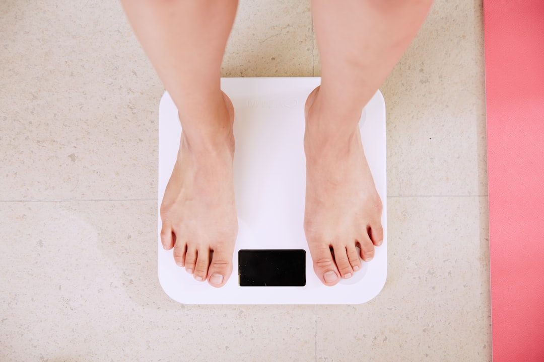
The result is a body composition change that the scale almost never captures accurately. A woman can weigh exactly the same as she did three years ago while simultaneously holding significantly less muscle and significantly more visceral fat. The number on the scale is unchanged. Her risk profile is not.
The Measurement Gap Nobody Talks About
Here is where the fitness and medical industries both fall short: they're measuring the wrong things.
Weight tells you nothing about the ratio of muscle to fat. BMI—derived entirely from weight and height—tells you even less. Body fat percentage estimates from handheld bioelectrical impedance devices are notoriously inaccurate and inconsistent, shifting by several percentage points based on hydration status, time of day, and how recently you ate. Even most primary care physicians don't routinely screen for muscle mass or visceral fat during annual physicals—not because they don't care, but because the standard diagnostic toolkit doesn't include those measurements.
If you're looking for an alternative to a DEXA scan, the honest answer is that there isn't one that comes close for soft tissue measurement. DEXA—dual-energy X-ray absorptiometry—is the clinical gold standard for measuring lean mass, fat mass, bone mineral density, and regional body composition simultaneously. It differentiates between subcutaneous fat (under the skin) and visceral fat (around the organs). It quantifies muscle mass by limb, identifying asymmetries that would otherwise be invisible. And it does all of this in under fifteen minutes.
For women in perimenopause, DEXA scanning doesn't just provide a number—it provides a baseline, a trajectory, and an early warning system. The women who come in once and never return learn something. The women who come in every sixty to ninety days learn something actionable.
What Perimenopause Muscle Weakness Actually Looks Like on a Scan
Perimenopause muscle weakness often doesn't announce itself as weakness first. It announces itself as effort. A workout that used to feel manageable starts to feel harder. A run that used to take forty-five minutes starts taking fifty-three. A set of squats that used to be done with a certain weight now requires two fewer reps to complete cleanly.
Women frequently attribute this to stress, poor sleep, or aging in general. All of those factors play a role. But the underlying mechanism—declining lean mass and reduced neuromuscular efficiency—is measurable before the subjective experience becomes undeniable.
On a DEXA scan, this shows up as a reduction in appendicular lean mass index (ALMI): the ratio of limb muscle mass to height squared. ALMI is one of the most clinically validated markers for sarcopenia risk, and it can begin declining meaningfully in a woman's early-to-mid forties—well before she meets any traditional diagnostic threshold for sarcopenia. As explored in tracking sarcopenia risk before symptoms appear, catching this decline early is the entire point.
DEXA also reveals regional asymmetries. A woman who favors one side during exercise, or who has a history of injury on one leg or shoulder, will often show measurably more lean mass on one side than the other. These imbalances matter for injury risk, movement quality, and long-term joint health—and they're invisible without regional data.
The Visceral Fat Problem Hidden Inside the Muscle Problem
Perimenopause doesn't just affect muscle—it changes where fat lives. As discussed in detail in why perimenopause belly fat is different, the hormonal environment of this transition actively promotes visceral fat accumulation. This matters because visceral fat is metabolically active in ways that subcutaneous fat is not. It secretes inflammatory cytokines, disrupts insulin signaling, and independently increases cardiovascular risk.
The troubling pattern Kalos sees repeatedly in perimenopausal women: they come in focused entirely on weight or body fat percentage, and the scan reveals a visceral fat score that's higher than expected given their overall body composition. They look lean. Their scale weight is within range. But the fat that actually drives disease risk has been accumulating around their organs, undetected.
This is the description problem the fitness industry hasn't solved. You can have 10,000 steps per day logged on your wearable, a resting heart rate that looks great, and sleep scores in the green—and still be developing a body composition profile that predicts poor metabolic health twenty years from now. The data exists to catch this. It's just not the data most people are looking at.
Why Tracking Intervals Matter More Than a Single Snapshot
A single DEXA scan is useful. Sequential scans are transformative.
The value of baseline measurement is that it gives you something to compare against. But the value of periodic scanning is that it catches trends—and in perimenopause, trends move in predictable directions if they're not actively interrupted.
Research consistently shows that women who do not engage in structured resistance training during perimenopause lose lean mass at an accelerated rate. Women who do engage in resistance training can preserve—and in many cases build—meaningful muscle during this window. But without measurement, you cannot confirm which category you're in. You might be training consistently and still losing muscle if your caloric intake is insufficient, your protein targets are too low, or your program isn't providing enough mechanical stimulus to offset the hormonal headwinds.
As covered in body recomposition after 45, effort and outcome don't always align the way we expect. A woman can do everything right subjectively—show up to the gym, eat well, manage stress—and still be losing ground on lean mass if the specific inputs aren't calibrated to her physiology. Measurement closes that gap.
This is why Kalos structures its coaching model around monthly scans. It's not about obsessing over numbers—it's about closing the feedback loop between what you're doing and what's actually happening in your body. If you're building lean mass and reducing visceral fat, the plan is working and you continue. If you're not, you adjust before months of effort are wasted in the wrong direction.
What Kalos Measures and Why It Matters for Perimenopausal Women
Every Kalos DEXA scan includes the metrics that matter most for women in this life stage:
Appendicular Lean Mass Index (ALMI): The clinical marker for sarcopenia risk. Kalos tracks this against age-matched reference populations so you know exactly where you stand relative to women your age—and how fast that's changing.
Visceral Adipose Tissue (VAT): Quantified in grams and translated into a risk score. This is the fat your doctor probably isn't measuring during your annual physical.
Bone Mineral Density (BMD): Estrogen decline accelerates bone loss, and the perimenopausal window is when intervention has the highest impact. Bone density declines silently—DEXA catches it before it becomes fracture risk.
Regional lean mass symmetry: Right arm versus left, right leg versus left. Asymmetries above certain thresholds predict injury risk and signal compensatory movement patterns worth addressing.
Total and segmental fat mass: Including the breakdown between subcutaneous and visceral, trunk versus peripheral—a distribution map that tells a more complete story than any single number.
The in-person analysis that follows every scan is where the data becomes a plan. Kalos coaches are NASM-certified and trained to translate raw scan output into specific, prioritized recommendations: what to change in your training, what to adjust in your nutrition, and what to monitor most closely at your next scan.
The Prescription Problem: Knowing What to Do With the Data
One of the two core problems Kalos was built to solve is the prescription problem: even when women have good data, they often don't know what to do with it. A DEXA scan report is a dense document filled with numbers, percentiles, and clinical terminology. Without interpretation, it's interesting but not actionable.
Kalos connects the dots between your DEXA metrics (the outcomes, or Y variables) and your behaviors (the inputs, or X variables). If your ALMI is declining, we look at resistance training volume and protein intake first—because those are the 80% inputs that drive lean mass outcomes. If visceral fat is rising despite consistent exercise, we examine nutrition quality and caloric context, because cardio alone doesn't reliably move visceral fat.
The goal isn't to prescribe a methodology and hope it works. The goal is to measure your response, observe what's actually moving, and adjust accordingly. Perimenopause doesn't respond to generic programs—it responds to precise, individualized intervention guided by real data.
The Window Is Open. It Doesn't Stay Open.
The perimenopausal transition is a window, not a permanent state. The hormonal environment that makes muscle loss and fat redistribution most likely to accelerate is also the environment in which targeted intervention has the highest return. Women who build muscle and maintain bone density during perimenopause enter postmenopause with a significantly stronger foundation—one that's measurably harder to build from scratch afterward.
The women who come into Kalos at 44 or 47 and start tracking with intention are not the same as the women who wait until 55 and then try to reverse a decade of undocumented decline. The biology is different. The opportunity is different. And the data, if collected, will prove it either way.
If you're in the Bay Area—San Francisco, Palo Alto, or San Jose—and you're in perimenopause or approaching it, a DEXA scan at Kalos is the most information-dense, clinically relevant thing you can do for your health right now. All services are HSA/FSA eligible.

Ready to measure what matters?
Book your DEXA scan today and stop guessing about your health.