Menopause Weight Gain: Is Fat Redistributing or Actually Increasing?

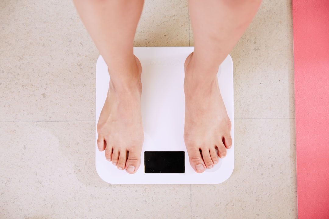
You step on the scale. The number is up—maybe five pounds, maybe ten. You haven't changed what you eat. You haven't stopped working out. But something is clearly different. Clothes fit differently. Your midsection looks different. You feel different.
The standard explanation you've probably heard: menopause slows your metabolism and makes you gain weight. Simple cause, simple effect.
Except it's not that simple. And for many women going through menopause, the real story is more complicated—and more actionable—than the scale suggests.
The question worth asking isn't just how much weight you've gained. It's whether you're actually gaining fat, losing muscle, or simply watching fat redistribute from one region of your body to another. These are three very different physiological events with three very different implications for your health, and no bathroom scale can tell them apart.
What Actually Happens to Body Composition During Menopause
Estrogen does a lot of things in the female body. One of its less-discussed roles is fat distribution regulation. Estrogen tends to promote fat storage in subcutaneous depots—the hips, thighs, and glutes. This is why premenopausal women typically carry more fat in those areas compared to men of similar age.
When estrogen levels drop during perimenopause and menopause, that regulatory signal weakens. Fat doesn't disappear. It migrates. The body shifts fat storage preference toward the abdomen—specifically toward visceral adipose tissue (VAT), the fat that accumulates around internal organs. This is the fat that carries meaningful metabolic and cardiovascular risk.
At the same time, declining estrogen and progesterone levels affect muscle protein synthesis. The hormonal environment that helped maintain lean mass through your 30s becomes less supportive. Without active intervention, muscle loss accelerates—sometimes quietly enough that nothing changes on the scale, but everything changes in body composition.
So when a woman in her late 40s or early 50s says "I've only gained two pounds but I look completely different," she's often describing something real: fat has redistributed from low-risk subcutaneous depots to high-risk visceral storage, while muscle has quietly declined. The scale caught almost none of it.
Three Scenarios the Scale Can't Distinguish
When the number on the scale goes up during menopause, at least three different things could be happening—or some combination of all three:
Scenario 1: Fat is redistributing, not increasing. Total fat mass stays roughly the same, but the location changes. Subcutaneous fat in peripheral regions decreases while visceral fat in the abdominal cavity increases. Weight on the scale may be unchanged or minimally different, but health risk increases substantially. You can't see visceral fat in the mirror. You can't feel it. Only clinical imaging can measure it.
Scenario 2: Fat is actually increasing. Total fat mass is genuinely going up—often because metabolic rate has declined, activity has decreased, or caloric intake hasn't adjusted to match a changing metabolism. This shows up on the scale, but the scale still can't tell you where the fat is going or what it's replacing.
Scenario 3: Muscle is decreasing while fat increases. This is the most clinically concerning scenario and the most commonly missed one. When muscle mass drops and fat mass rises simultaneously, the scale can show almost no change—or even a decrease—while body composition deteriorates significantly. A woman might lose four pounds of muscle and gain three pounds of fat and conclude from the scale that things are "fine." They're not.
Each of these scenarios calls for a different response. Redistribution without total gain might prioritize visceral fat reduction through targeted exercise and stress management. Genuine fat gain calls for a recalibration of nutrition and energy balance. Muscle loss demands a protein and resistance training protocol, urgently. Treating all three the same way—just "eating less and moving more"—is why so many women feel like they're doing everything right and seeing nothing change.
Why This Matters More Than the Number on the Scale
Visceral fat isn't just aesthetically different from subcutaneous fat. It's metabolically different. VAT releases inflammatory cytokines, disrupts insulin signaling, and is associated with elevated cardiovascular risk, type 2 diabetes, and metabolic syndrome. A woman who has gained no weight but has experienced significant visceral fat accumulation is at meaningfully higher risk than her scale suggests.
Muscle mass matters for the opposite reasons. Skeletal muscle is metabolically active tissue—it burns calories at rest, supports insulin sensitivity, and protects against sarcopenia (age-related muscle loss) that becomes clinically significant in later decades. As we've explored in why Bay Area professionals are using DEXA scans to measure whether menopause is causing hidden muscle loss, the muscle loss that happens during this transition often goes entirely undetected until symptoms become hard to ignore.
The concern isn't vanity. It's function. It's longevity. It's the difference between a body that remains resilient and strong through your 60s and 70s versus one that becomes increasingly fragile in ways that compound over time. Perimenopause belly fat is physiologically distinct—and understanding that distinction early is what allows you to respond appropriately.
What DEXA Scanning Actually Measures
A DEXA (dual-energy X-ray absorptiometry) scan is the clinical gold standard for body composition measurement. It uses two low-dose X-ray beams at different energy levels to differentiate between bone, lean tissue, and fat tissue with a level of precision that no other widely available method can match.
In a single 10-minute scan, you get a precise breakdown of:
- Total fat mass — the absolute amount of fat in your body
- Regional fat distribution — how much fat is in your trunk versus arms versus legs
- Visceral adipose tissue (VAT) — the specific fat surrounding your internal organs, quantified as a discrete metric with established risk thresholds
- Lean mass by region — how much muscle you have in each limb and your trunk, and whether it's symmetric
- Bone mineral density (BMD) — critically important during and after menopause, when estrogen loss accelerates bone resorption
- Appendicular lean mass index (ALMI) — a standardized measure of muscle relative to height that predicts sarcopenia risk
This is the difference between knowing your total weight and understanding your body. When someone asks where to get a DEXA scan near me in the Bay Area, what they're often really asking is: how do I finally get real data about what's happening inside my body?
A DEXA scan answers the redistribution question directly. If your VAT has increased by 200 grams since your last scan while your subcutaneous trunk fat has stayed the same, that's redistribution. If your total fat mass has increased by two kilograms concentrated in the abdominal region, that's accumulation. If your lean mass in your legs has dropped by half a kilogram over six months, that's muscle loss—caught before it compounds further. These are three completely different clinical pictures requiring three completely different interventions.
The Menopause Body Shape Conversation Nobody Is Having
There's a lot of conversation in the wellness space about menopause body shape changes—the shift from a pear to an apple, the loss of waist definition, the belly that "won't go away no matter what." Most of this conversation stops at observation. It describes the change without quantifying it, and without that quantification, it's nearly impossible to respond effectively.
Knowing that you've shifted toward more central fat storage is qualitatively useful. Knowing that your VAT score has crossed from a low-risk to a moderate-risk threshold—and by exactly how much—is actionable. Knowing that your appendicular lean mass index places you at early sarcopenia risk means you can start a resistance protocol now, before the losses compound. Tracking muscle loss at regular intervals is how you turn a vague concern into a precise intervention target.
This is also where the distinction between redistribution and accumulation becomes practically critical. A woman experiencing primarily redistribution might not need a large caloric deficit—she might need a visceral fat reduction protocol that emphasizes high-intensity interval work, stress management, and sleep optimization. A woman experiencing genuine fat accumulation alongside muscle loss needs a carefully constructed protein-forward nutrition strategy combined with progressive resistance training. These are not the same prescription. Getting the wrong one doesn't just fail to help—it can make things worse.
What Happens When You Only Track the Scale
Consider the pattern that plays out repeatedly: a woman in her late 40s notices her body changing. She cuts calories. She exercises more, often adding cardio. The scale drops slightly or stays flat. She concludes the intervention is working—or that it's "just menopause" and nothing can be done.
Meanwhile, without DEXA data, she has no idea whether she's losing muscle along with fat. She has no idea whether her visceral fat is actually responding to the cardio, or whether it's accumulating despite her efforts. She has no idea if her bone density is declining—a silent process that DEXA catches years before fracture risk becomes apparent. As we've seen in data from postmenopausal women regaining weight, the picture beneath the surface is almost always more complex than the scale suggests.
Six months of effort without the right measurement layer is six months of guessing. Body recomposition after 45 is absolutely achievable—but only when the effort is matched to what the data is actually showing, not what the scale implies.
Bone Density: The Third Variable Most Women Miss
Estrogen plays a protective role in bone metabolism. As estrogen declines during perimenopause and menopause, bone resorption accelerates. The clinical risk of osteoporosis increases substantially in the decade following menopause, and the loss often begins years before a woman or her doctor would think to check.
A standard annual physical doesn't include bone density assessment for most women until age 65, by which point significant loss may have already occurred. DEXA scanning catches this early. The bone density loss that begins during perimenopause can be identified, tracked, and meaningfully addressed through resistance training and targeted nutrition—but only if you know it's happening.
This matters in the context of menopause body composition tracking because fat, muscle, and bone are not independent variables. They are part of an interconnected system. The hormonal changes that shift fat distribution also affect muscle synthesis and bone resorption simultaneously. Measuring only one piece of this system gives you an incomplete—and potentially misleading—picture.
How Kalos Approaches Menopause Body Composition
At Kalos, DEXA scanning is the measurement layer. It's not the product—it's the foundation on which everything else is built. When a woman comes in during perimenopause or post-menopause with concerns about body composition changes, a baseline DEXA scan gives us exact numbers across every dimension that matters: total fat, regional fat, VAT, lean mass by region, ALMI, and bone density.
That data is then interpreted in a coaching session by a NASM-certified performance analyst who specializes in connecting body composition metrics to actionable protocols. We're not handing you a printout and wishing you luck. We're using the data to answer: what specifically is changing in your body, why is it changing, and what do you do about it?
For our longevity-focused members, this framing resonates deeply. Optimizing for longevity means protecting bone density now, preserving muscle mass aggressively, and keeping visceral fat within healthy bounds—not just chasing a number on a scale. For members tracking perimenopause changes, tracking muscle shifts during perimenopause through serial DEXA scans turns a confusing and often demoralizing experience into a clear, data-driven process.
We also use Kalos's proprietary Triangle Framework to orient every coaching conversation. Every member's goals sit somewhere on a triangle connecting Aesthetics, Longevity, and Performance. For most women navigating menopause body composition changes, longevity metrics—VAT, BMD, ALMI—are the primary targets. But improving these metrics almost always improves aesthetics and performance outcomes simultaneously. The trade-offs only appear at the extremes. For most people, the data points in the same direction across all three vertices.
Practical Steps for Getting Real Answers
If you're in the Bay Area and experiencing menopause-related body changes, here's the sequence that actually produces clarity:
Step 1: Get a baseline DEXA scan. Stop guessing what's happening. A 10-minute scan at Kalos (San Francisco, Palo Alto, or San Jose) gives you exact fat mass, lean mass, VAT, and bone density data. This is the only way to answer the redistribution-versus-accumulation question with precision.
Step 2: Interpret the data with a coach. A scan alone is a starting point, not a prescription. Your performance analyst reviews the numbers with you and identifies which metrics are most concerning, which are most actionable, and which interventions are most likely to move your specific numbers. This is the prescription problem the fitness industry consistently fails to solve.
Step 3: Build a protocol grounded in your actual data. Resistance training prioritized for muscle preservation and bone density. Nutrition calibrated around protein targets that support lean mass retention. Cardiovascular work chosen for its effect on VAT specifically. These aren't generic recommendations—they're derived from what your scan actually shows.
Step 4: Rescan at regular intervals. Body composition is not a one-time measurement. It's a time series. Monthly or quarterly rescans tell you whether the protocol is working—whether muscle is being preserved, whether VAT is responding, whether bone density is holding. Retesting after 60 days is often enough to see clear directional signals. If the approach isn't working, the data tells you that before months of effort go to waste.
The Bottom Line
Menopause body changes are real, significant, and worth taking seriously. But "weight gain" is too blunt a description for what's actually happening. Some of it is redistribution—the same fat showing up in a more dangerous place. Some of it is genuine accumulation. Some of it is muscle loss masquerading as stable weight. Most women are experiencing all three simultaneously, in varying proportions, with no tool sensitive enough to tell them which is which.
The scale is not that tool. BMI is not that tool. Even a well-intentioned primary care visit is unlikely to produce the granular body composition data that makes the difference between an effective intervention and a frustrating guess.
A DEXA scan is that tool. And in the Bay Area, Kalos is where you come to use it—and to turn what it reveals into a plan that actually works.
All services at Kalos are HSA/FSA eligible. We have locations in San Francisco, Palo Alto, and San Jose. If you're ready to stop guessing and start measuring, your baseline scan is the first step.

Ready to measure what matters?
Book your DEXA scan today and stop guessing about your health.