Bone Density Testing: DEXA Scans Versus Other Methods Compared
Why Bone Density Testing Is Getting More Attention in 2026
Bone density used to be a conversation that started at a primary care appointment sometime after 65. That's changing fast. Bay Area professionals in their late 30s and 40s—particularly those who are active, health-conscious, and already tracking other biomarkers—are increasingly asking a question their annual physical never raises: What is actually happening inside my bones right now?
The interest is well-founded. Bone mineral density (BMD) is one of the most predictive longevity metrics available. Declining BMD is largely silent until a fracture occurs. And while the popular image of osteoporosis is an elderly woman with a stooped posture, the bone loss that causes it begins in midlife—sometimes earlier, particularly in women approaching perimenopause and in men with low testosterone or high cortisol loads.
If you've been searching for "dexa bone density scan near me" or "where to get bone density test near me," you've likely discovered that the options are more varied than you expected. Hospital radiology departments, independent DEXA facilities, portable ultrasound devices, and even some primary care offices all claim to offer bone density testing. The differences between these methods are significant—and choosing the wrong one can leave you with data that's technically a number but practically meaningless.
This guide breaks down every major bone density testing method available in 2026, compares them honestly, and explains why the method you choose should depend on what you're actually trying to learn.
What Bone Density Testing Actually Measures
Before comparing methods, it's worth being precise about what bone density testing measures—because "bone density" is used loosely in ways that obscure important distinctions.
Bone mineral density (BMD) refers to the concentration of minerals, primarily calcium, within a given area of bone tissue. Higher mineral density generally indicates stronger, more fracture-resistant bone. Lower density—classified clinically as osteopenia or osteoporosis using T-scores—indicates elevated fracture risk.
Results are typically expressed as a T-score, which compares your BMD to the average peak bone mass of a healthy young adult of the same sex. A T-score of 0 is average. A T-score of -1.0 to -2.5 indicates osteopenia. Below -2.5 indicates osteoporosis. A Z-score, less commonly used clinically, compares your BMD to age-matched peers rather than young adult norms.
Most bone density tests focus on two sites: the lumbar spine and the proximal femur (hip). These sites are prioritized because they are the most clinically significant locations for fracture risk, and because they contain trabecular bone—a porous, metabolically active bone type that responds more rapidly to changes in nutrition, exercise, hormones, and aging than cortical (compact) bone does.
With that foundation established, here is a direct comparison of every method currently available.
Method 1: DEXA (Dual-Energy X-Ray Absorptiometry)
What It Is
DEXA uses two low-dose X-ray beams at different energy levels to distinguish bone from soft tissue. By measuring how much each beam is attenuated as it passes through the body, the system calculates mineral content and density at any measured site. A full-body DEXA scan takes 10 to 20 minutes, involves no preparation beyond removing metal objects, and exposes the subject to roughly 1–10 microsieverts of radiation—less than a day of natural background radiation, and a small fraction of a chest X-ray.
What It Measures
A clinical DEXA scan measures BMD at the lumbar spine and femoral neck in detail. A full-body DEXA scan—the type used at performance health facilities like Kalos—simultaneously captures BMD at multiple sites alongside a complete body composition picture: total and regional fat mass, lean mass (muscle), visceral adipose tissue (VAT), and the Appendicular Lean Mass Index (ALMI), which is the gold-standard diagnostic marker for sarcopenia.
This is not a minor distinction. When you get a DEXA scan for bone density, you are also getting your body fat percentage measured to clinical accuracy, your muscle mass broken down by limb and region, your visceral fat level quantified, and your lean mass index benchmarked against age and sex norms. No other bone density testing method comes close to this breadth of information from a single test.
Accuracy and Clinical Standing
DEXA is the gold standard for bone density measurement. It is the method used in virtually all clinical trials studying BMD. It is the method recommended by the International Society for Clinical Densitometry (ISCD), the National Osteoporosis Foundation, and the World Health Organization. The T-score thresholds that define osteopenia and osteoporosis were developed using DEXA data. When other methods advertise accuracy, they are comparing themselves to DEXA as the reference standard.
Precision error—the variability you'd see if you scanned the same person twice on the same day—is approximately 1–2% for spine and hip BMD on modern DEXA equipment. This precision is critical for longitudinal tracking: it means that if your BMD changes by 3–4%, the scan can detect that change as real rather than noise.
Limitations
DEXA is not portable. It requires a fixed machine operated by trained technicians. Results can be affected by positioning errors, patient movement, artifacts from metal implants, and the specific software and calibration of the machine. For this reason, tracking BMD changes over time is most accurate when scans are performed on the same machine at the same facility. Cross-facility comparisons require careful calibration adjustments.
DEXA also does not differentiate between cortical and trabecular bone compartments the way some more advanced methods do—a limitation that matters primarily in research contexts rather than clinical or performance health applications.
Availability and Cost
DEXA is widely available in the Bay Area through hospital radiology departments, independent imaging centers, and performance health facilities. Costs range from a one-time scan fee to inclusion in a coaching membership. At Kalos, DEXA scanning serves as the measurement foundation for a broader performance health transformation process—not just a single data point.
Method 2: Quantitative Computed Tomography (QCT)
What It Is
QCT uses a standard CT scanner to generate three-dimensional images of bone and calculate volumetric BMD (vBMD) in grams per cubic centimeter, rather than the areal BMD (aBMD) in grams per square centimeter that DEXA measures. QCT can be performed on the spine (central QCT) or peripheral sites like the forearm or tibia (peripheral QCT, pQCT).
What It Measures
QCT's three-dimensional approach allows it to separately measure trabecular and cortical bone compartments, which DEXA cannot do directly. This is its primary clinical advantage—it can detect early trabecular bone loss before it becomes apparent on DEXA, making it potentially useful for early-stage monitoring in high-risk populations.
Accuracy and Clinical Standing
QCT is highly accurate and is used in research settings. However, it has not replaced DEXA as the clinical standard because the T-score thresholds for diagnosing osteoporosis were established using DEXA data, not QCT data. QCT results are not directly comparable to DEXA T-scores, which limits their clinical interpretability in standard practice.
Limitations
QCT involves significantly higher radiation exposure than DEXA—roughly 50 to 100 times more for a spinal scan, though peripheral QCT involves far less. It is more expensive, less widely available, and lacks standardized diagnostic thresholds for most clinical decisions. For these reasons, it is rarely used for routine bone density assessment outside of research or highly specialized clinical contexts.
Verdict for Most People
QCT is a powerful research tool but not a practical choice for routine bone density monitoring. Its advantages over DEXA in separating trabecular from cortical bone are meaningful in specific research and high-risk clinical scenarios—not in the performance health context where most Bay Area professionals are operating.
Method 3: Quantitative Ultrasound (QUS)
What It Is
Quantitative ultrasound uses sound waves—not radiation—to assess bone properties, typically at the heel (calcaneus), though some devices measure the finger, wrist, or tibia. It measures two parameters: Broadband Ultrasound Attenuation (BUA), which reflects bone density, and Speed of Sound (SOS), which reflects bone elasticity. These are combined into a composite score called Stiffness Index or Quantitative Ultrasound Index (QUI).
What It Measures
QUS at the heel provides information about bone quality at a peripheral site. It does not directly measure BMD at the spine or hip—the sites most relevant to clinical fracture risk and the sites where DEXA's T-scores are actually defined. A heel ultrasound result cannot be used to diagnose osteoporosis by WHO criteria.
Accuracy and Clinical Standing
QUS has been studied extensively as a fracture risk screening tool. It shows reasonable correlation with hip fracture risk in older women—meaning populations with low QUS scores do tend to have higher fracture rates. However, it cannot replace DEXA for diagnosis. Most clinical guidelines, including those from the ISCD, state explicitly that QUS results cannot be used to diagnose osteoporosis or monitor treatment response.
Limitations
QUS cannot diagnose osteoporosis. It cannot monitor the effects of bone-protective medications or interventions. It measures a peripheral site (the heel) rather than the spine and hip where clinical decisions are made. Results vary significantly across device brands, with no universal reference standard. Soft tissue effects, foot positioning, and the temperature of the coupling gel all affect results.
QUS is sometimes presented as a screening tool—a way to identify people who should then go get a DEXA scan. In that limited role, it has some utility. As a standalone bone health assessment, it is inadequate.
Verdict for Most People
Quantitative ultrasound is a low-cost, no-radiation option that can prompt someone to pursue further testing. It is not a replacement for DEXA and should not be treated as one. If you're searching for "where to get bone density test near me" and the answer involves a heel ultrasound at a pharmacy kiosk or health fair, recognize that this is a screening prompt—not a diagnosis and not a trackable baseline.
Method 4: Peripheral DEXA (pDEXA)
What It Is
Peripheral DEXA uses the same dual-energy X-ray technology as central DEXA but applies it to peripheral sites: typically the wrist, heel, or finger. These devices are smaller, less expensive, and more portable than central DEXA machines.
What It Measures
pDEXA measures BMD at peripheral cortical bone sites. Like QUS, it does not measure spine or hip BMD. It can identify low BMD at a peripheral site, which may correlate with low BMD elsewhere, but it cannot be used to diagnose osteoporosis by standard criteria or to make treatment decisions.
Accuracy and Clinical Standing
pDEXA has better standardization than QUS, but it still cannot substitute for central DEXA in clinical decision-making. Peripheral BMD at the wrist does not predict spine or hip fracture risk with the same reliability as central measurements.
Limitations
The same core limitation as QUS: it measures the wrong sites for clinical decision-making. It is sometimes used in community screening programs to identify people who should receive a central DEXA scan.
Verdict for Most People
Peripheral DEXA is more reliable than QUS but still not a substitute for central DEXA. Treat it the same way: as a screening prompt, not a complete assessment.
Method 5: High-Resolution Peripheral Quantitative CT (HR-pQCT)
What It Is
HR-pQCT is an advanced imaging technique that produces extremely high-resolution three-dimensional images of peripheral bone microarchitecture—typically at the distal radius (forearm) and tibia. It can assess trabecular number, thickness, and separation; cortical thickness and porosity; and estimated bone strength through finite element analysis.
What It Measures
HR-pQCT measures bone quality at a microstructural level in ways that no other technique can. It is the most detailed bone assessment tool available in clinical research.
Accuracy and Clinical Standing
Highly accurate for microarchitectural assessment. Used in academic medical centers and research institutions. Not used in routine clinical practice due to cost, limited availability, and the absence of standardized diagnostic thresholds for clinical decisions.
Limitations
Unavailable in most non-academic settings. No established diagnostic T-score equivalent. Not used to guide clinical treatment decisions. Radiation exposure, while lower than QCT, is higher than DEXA. Practically speaking, this method is a research tool, not a performance health tool.
Verdict for Most People
Unless you are enrolled in a research study or being seen at a specialized academic medical center, HR-pQCT is not relevant to your bone density assessment. File it under "interesting science, not available to you."
Method 6: Radiographic Absorptiometry and Other Legacy Methods
Several older methods—radiographic absorptiometry, single-energy X-ray absorptiometry (SXA), and single-photon absorptiometry (SPA)—were used before DEXA became the standard. You may encounter references to them in older literature. They have been superseded by DEXA and are rarely used clinically today. They are included here only for completeness; they are not worth pursuing in 2026.
Side-by-Side Comparison: What Actually Matters
Here is a direct comparison across the dimensions that matter most when choosing a bone density testing method:
Measures spine and hip BMD directly: Central DEXA — Yes. QCT — Yes. QUS — No. pDEXA — No. HR-pQCT — No (peripheral only).
Can diagnose osteoporosis by WHO criteria: Central DEXA — Yes. QCT — Partially (no standardized T-scores). QUS — No. pDEXA — No. HR-pQCT — No.
Can track changes over time: Central DEXA — Yes. QCT — Yes (research settings). QUS — Poorly. pDEXA — Poorly. HR-pQCT — Yes (research).
Includes full body composition: Central DEXA — Yes. All others — No.
Radiation involved: Central DEXA — Minimal (1–10 µSv). QCT — Moderate to high. QUS — None. pDEXA — Minimal. HR-pQCT — Low to moderate.
Widely available in Bay Area: Central DEXA — Yes. QCT — Yes (hospitals). QUS — Yes (some pharmacies, health fairs). pDEXA — Limited. HR-pQCT — Academic centers only.
Appropriate for performance health tracking: Central DEXA — Yes. All others — No.
Why Bone Density Matters Beyond Fracture Prevention
The clinical framing of bone density testing—focused almost entirely on fracture risk and osteoporosis diagnosis—underserves the majority of people who should be paying attention to their BMD. For Bay Area professionals in their 30s, 40s, and 50s, the fracture conversation is premature. The performance and longevity conversation is not.
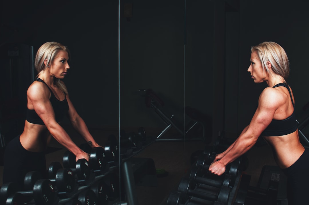
Bone mineral density is a meaningful longevity metric for several reasons beyond fracture prevention. Low BMD in midlife is associated with higher all-cause mortality independent of fracture events. BMD reflects cumulative hormonal health—particularly estrogen and testosterone levels over time—and serves as a proxy for hormonal function. BMD responds to resistance training in ways that make it a useful outcome metric for evaluating whether a strength training program is actually working at a structural level.
This is why Bay Area professionals are increasingly using DEXA scans to monitor bone density proactively—not waiting for a fracture or a physician referral, but treating BMD as one data point in a broader picture of how their body is aging and adapting.
For women in their 40s, the bone density conversation is particularly urgent. Perimenopause brings a dramatic acceleration in bone loss as estrogen levels decline—and this loss begins before the menstrual cycle becomes irregular. Research on perimenopause and bone density loss shows that the decade before menopause is a critical window for intervention. A DEXA scan taken during this period establishes a baseline that makes subsequent changes detectable and actionable.
For anyone on GLP-1 medications like Ozempic or Wegovy, bone density monitoring is especially important. Rapid weight loss is associated with bone loss—and the muscle loss that frequently accompanies GLP-1-driven weight reduction compounds this effect. Tracking the real effects of GLP-1 medications on body composition, including bone density, is one of the most clinically important reasons to get a DEXA scan in 2026.
The Full-Body DEXA Advantage: Bone Density Is Only the Beginning
When you come in for a DEXA scan at a performance health facility, you are not just getting a bone density number. You are getting a complete body composition picture that no other single test can provide.
In a single 10-to-20-minute scan, a full-body DEXA produces:
- Bone mineral density at the spine and hip, with T-scores and Z-scores
- Total body fat percentage, accurate to within 1–2%
- Regional fat distribution—arms, legs, trunk, android (belly), gynoid (hip/thigh)
- Visceral adipose tissue (VAT) quantification—the metabolically dangerous fat that wraps around organs and is invisible to any external measurement
- Lean mass (muscle) broken down by limb and region, allowing detection of asymmetry and imbalance
- Appendicular Lean Mass Index (ALMI), benchmarked against age and sex norms—the diagnostic marker for sarcopenia
Each of these metrics connects to a different dimension of the health triangle that shapes how we think about body composition at Kalos. Bone mineral density maps to longevity. Visceral fat maps to longevity and metabolic risk. Muscle mass maps to both performance and longevity. Body fat percentage maps to aesthetics and performance. Understanding visceral fat specifically is one of the most important things a DEXA scan can reveal—particularly for lean-looking professionals who have no obvious external signs of metabolic risk.
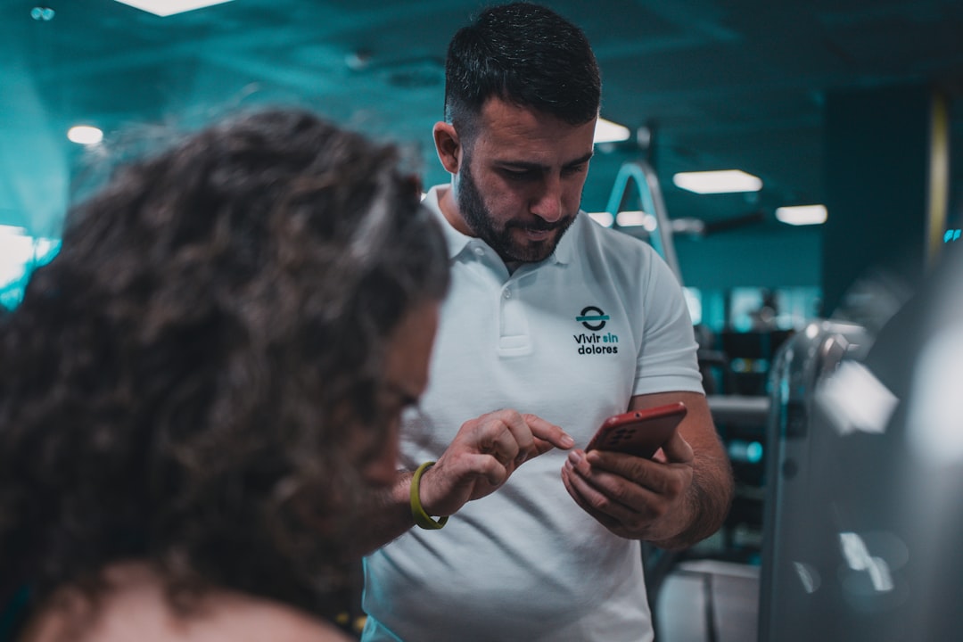
This is the core difference between a bone density test at a hospital radiology department and a DEXA scan at a performance health facility. At a hospital, you get a T-score and a one-page report that may or may not come with any clinical follow-up. At Kalos, your scan is analyzed by a NASM-certified performance analyst who walks you through every metric, explains what it means for your specific goals, and connects it to a personalized plan for exercise, nutrition, and ongoing tracking.
The measurement is the entry point. The transformation is the goal.
How Bone Density Fits Into a Broader Body Composition Assessment
One of the most common misunderstandings we see is treating bone density in isolation. A low BMD number does not tell you why your BMD is low or what to do about it. That requires context.
Is your lean mass adequate? Low muscle mass and low bone density frequently co-occur because both reflect insufficient mechanical loading and hormonal signaling. Improving resistance training volume often improves both simultaneously. DEXA-guided strength training optimization can help you design a program that specifically addresses bone loading, not just muscle growth.
Is your visceral fat elevated? High visceral fat is associated with chronic low-grade inflammation that impairs bone remodeling. Addressing VAT often improves multiple longevity markers together.
Are you losing weight rapidly—whether through dieting, GLP-1 medications, or high training volume? Rapid weight loss is one of the most reliable accelerants of bone loss. Tracking BMD during periods of deliberate caloric restriction is not optional—it is how you protect yourself from trading one problem for another. Muscle loss during dieting and bone loss during dieting are related problems with related solutions: sufficient protein, sufficient resistance training, and data-driven monitoring.
Are you a woman in perimenopause or approaching it? The hormonal shifts of this period directly suppress bone formation and accelerate resorption. A DEXA baseline taken at 40–45 is dramatically more useful than one taken at 55 after significant loss has already occurred.
Who Should Get a DEXA Scan for Bone Density—and When
Clinical guidelines for bone density testing are conservative—they generally recommend DEXA for women over 65 and men over 70, or for younger individuals with specific risk factors like glucocorticoid use, rheumatoid arthritis, or a prior fragility fracture. These guidelines are designed for clinical diagnosis and treatment decisions in a medical system focused on prevention of established disease.
Performance health operates on a different timeline. If you are waiting for clinical guidelines to tell you to get a bone density test, you are optimizing for the moment the problem becomes undeniable—not for the moment you can most effectively do something about it.
Consider getting a DEXA scan that includes bone density assessment if:
- You are a woman in your late 30s or 40s and have not established a BMD baseline
- You are currently on or considering GLP-1 medications and want to track the full effect on body composition, including bone
- You are experiencing perimenopausal symptoms and want to know whether bone loss has begun
- You have been in a sustained caloric deficit and want to confirm you are preserving bone mass alongside fat loss
- You have a family history of osteoporosis
- You want to establish a baseline before beginning a new resistance training program, so you can measure whether your bones are responding to the stimulus
- You are generally optimizing your healthspan and want a complete picture of how your body is aging
Finding Bone Density Testing Near You in the Bay Area
If you are in San Francisco, Palo Alto, or San Jose and searching for "bone density testing near me" or "dexa bone density scan near me," you have several categories of options:
Hospital radiology departments offer clinical DEXA scans that typically focus exclusively on spine and hip BMD. These are appropriate for clinical diagnosis but do not include body composition data, do not include coaching or analysis, and are usually ordered by a physician. Scheduling can take weeks.
Independent imaging centers offer DEXA scans without a physician referral in most cases. Results are typically limited to bone density data and may or may not include any interpretive guidance.
Performance health facilities like Kalos offer full-body DEXA scanning that simultaneously captures bone density and complete body composition—with in-person analysis from a certified performance analyst who connects your data to a specific, personalized action plan. HSA/FSA eligible. No physician referral required. Locations in San Francisco, Palo Alto, and San Jose.
The distinction matters. Bone density data without context is a number. Bone density data within a complete body composition picture, analyzed by someone who understands performance health and can connect it to your specific goals, is a foundation for change.
If you have been building a longevity stack and have not yet established your BMD baseline alongside your body composition baseline, the scan is where that work starts. The analysis is where it becomes actionable.
The Bottom Line
DEXA is the gold standard for bone density testing. Not because it is the most technologically sophisticated method available—QCT and HR-pQCT produce more detailed information in specific research contexts—but because it is the method against which all other methods are measured, the method used to establish the diagnostic thresholds the entire field relies on, and the only method that simultaneously captures a complete body composition picture alongside bone density data.
Quantitative ultrasound and peripheral DEXA are screening tools. They can tell you whether to go get a real scan. They cannot replace one.
QCT is a research tool with meaningful clinical limitations and substantially higher radiation exposure. HR-pQCT is a specialized research tool that is not practically available to most people.
If you are in the Bay Area and want to know your bone density with clinical-grade accuracy—and to understand it within the full context of your body composition, your muscle mass, your visceral fat, and your overall health trajectory—a full-body DEXA scan at Kalos is the right starting point.
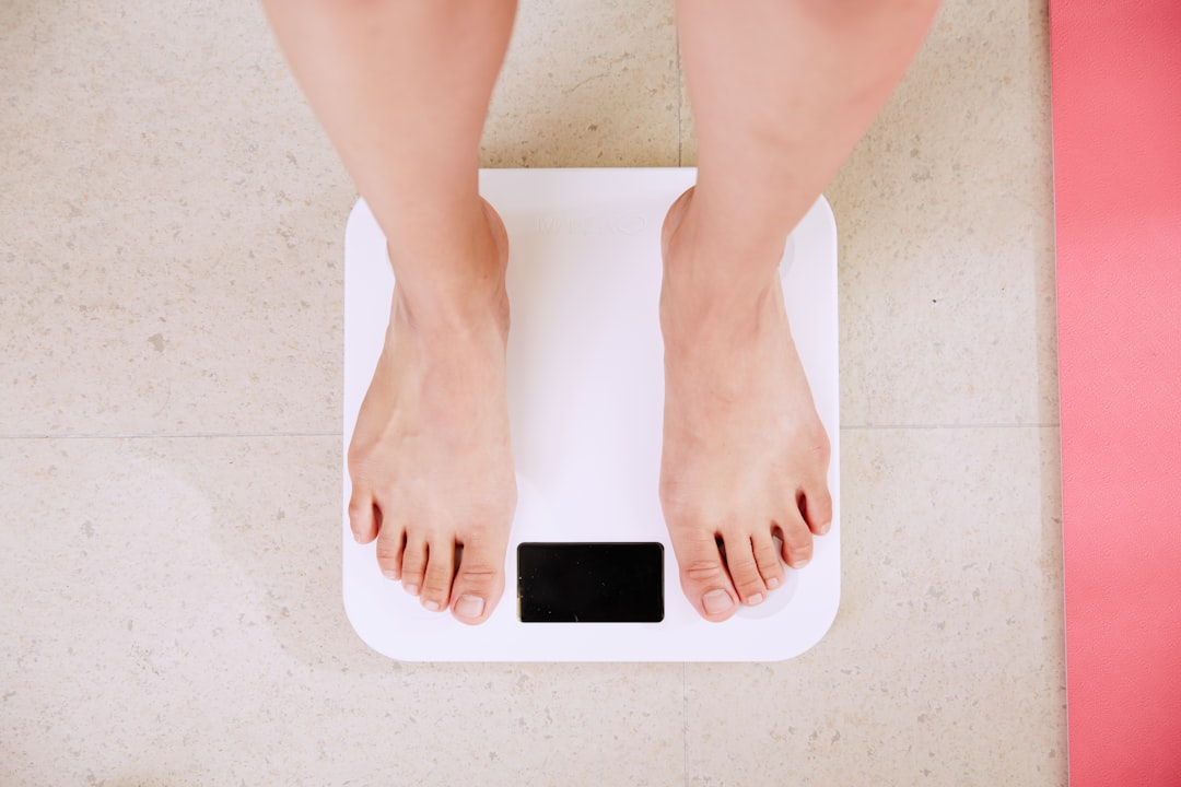
The measurement takes less than 20 minutes. The picture it gives you is one that your annual physical, your scale, your BMI, and your wearable cannot come close to providing.

Ready to measure what matters?
Book your DEXA scan today and stop guessing about your health.